This is an accessible summary of the peer-reviewed article, not a replacement for it. Read the source PDF for the complete methods, results, supporting-information references, and publication context.
Publication
Kennon, B. S.; Niedermeyer, W. H. “EVQ-218: Characterization of High-Energy Nanoparticles that Measure up to NIST Standards.” ACS Omega 2024, 9, 7891-7903. https://doi.org/10.1021/acsomega.3c07745
The article is published under a CC BY-NC-ND 4.0 license. This page summarizes its reported methods and findings in a more accessible web format.
Study overview
The study compares EVQ-218, a laser-ablated silver particle produced directly in high-purity water, with a NIST-traceable nanoComposix silver-particle reference. The authors evaluated particle morphology and size distribution, behavior in mineral-containing media, silver dissolution, surface chemistry, and long-term particle stability.
What the researchers tested
- Scanning transmission electron microscopy (STEM) for morphology and particle sizing.
- Dynamic light scattering (DLS) for particle-size distributions in different media.
- ICP-OES and a silver-ion-selective electrode for dissolution and silver measurements.
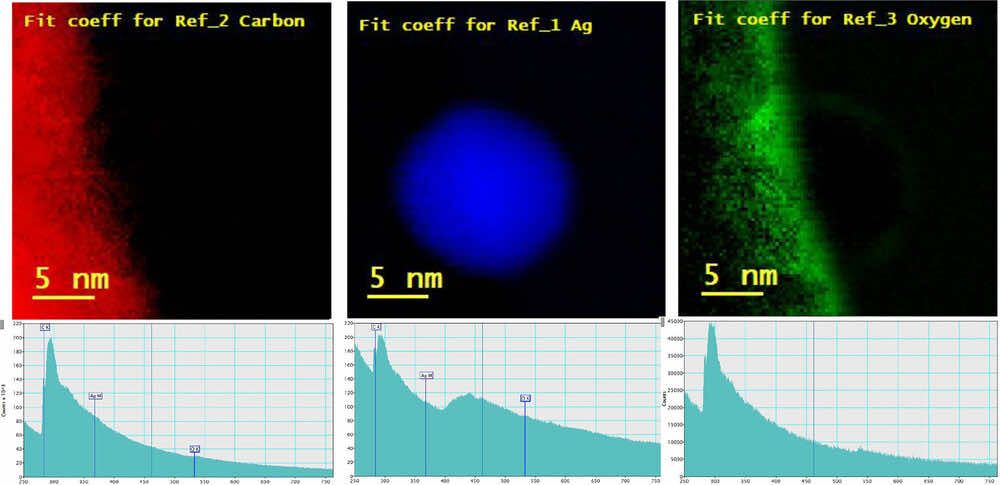
- STEM/EDS and EELS for elemental mapping and surface-chemistry characterization.
- Deionized water, moderately hard water, and concentrated artificial alveolar fluid as test media.
Particle morphology and sizing
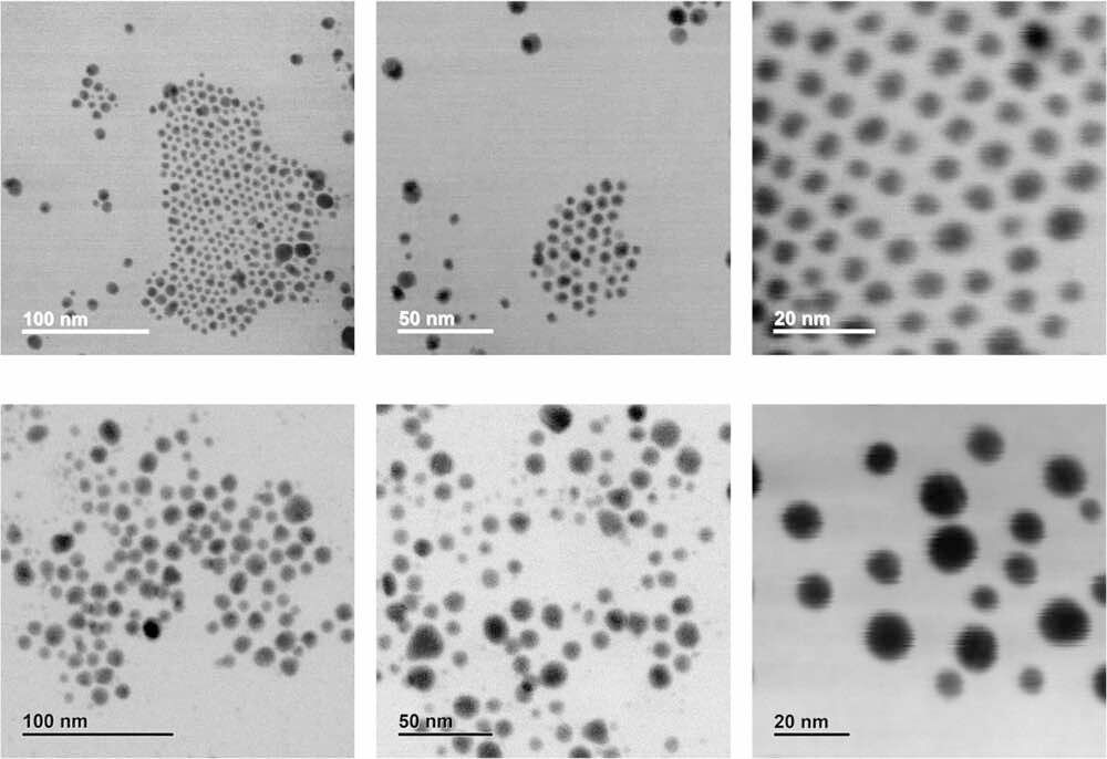
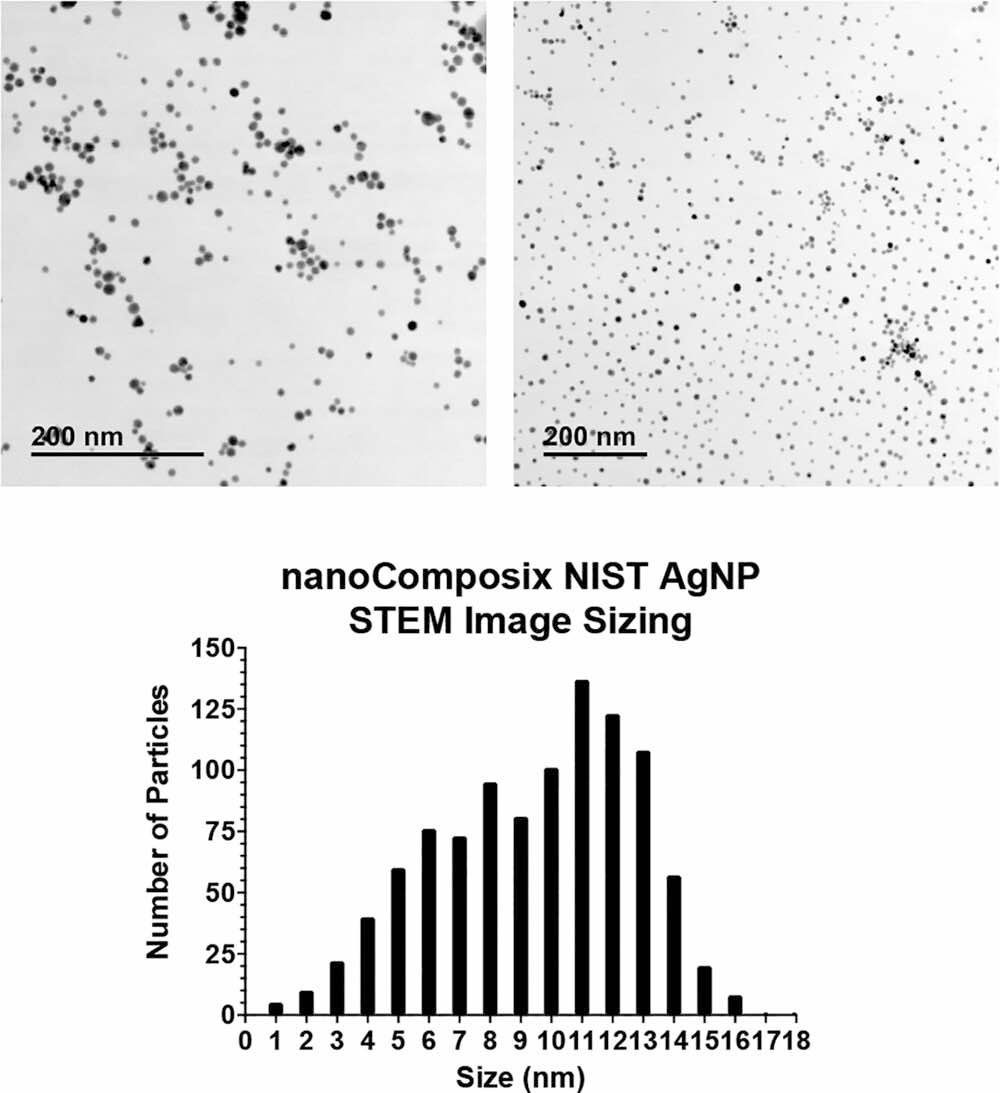
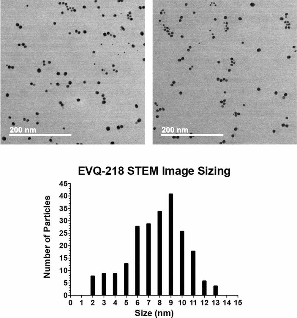
The authors report that EVQ-218 had a predominantly spherical morphology and a narrow particle-size distribution. Images showed small, regularly spaced groupings on the microscopy grid without the fused agglomeration that would indicate loss of particle identity. The NIST-traceable comparison material also had a narrow distribution, although the paper notes more faceted and nonspherical particles in the reference images.
Particle statistics reported in the article
| Percentile | EVQ-218 diameter | Number density | Specific surface area |
|---|---|---|---|
| d(10) | 3.9 nm | 3.2 × 10¹³ NP/mL | 148.5 m²/g |
| d(50) | 8.0 nm | 3.5 × 10¹² NP/mL | 71.1 m²/g |
| d(90) | 10.8 nm | 1.5 × 10¹² NP/mL | 53.1 m²/g |
| Percentile | NIST-reference diameter | Number density | Specific surface area |
|---|---|---|---|
| d(10) | 8.5 nm | 3.0 × 10¹² NP/mL | 67.5 m²/g |
| d(50) | 11.5 nm | 1.2 × 10¹² NP/mL | 49.9 m²/g |
| d(90) | 13.7 nm | 7.0 × 10¹¹ NP/mL | 41.6 m²/g |
Dissolution and behavior in test media
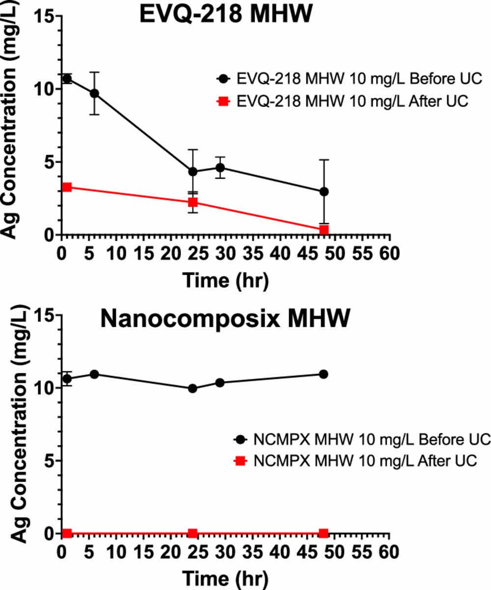
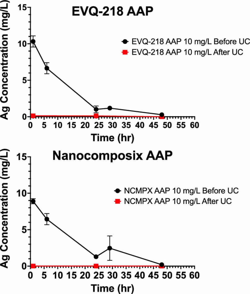
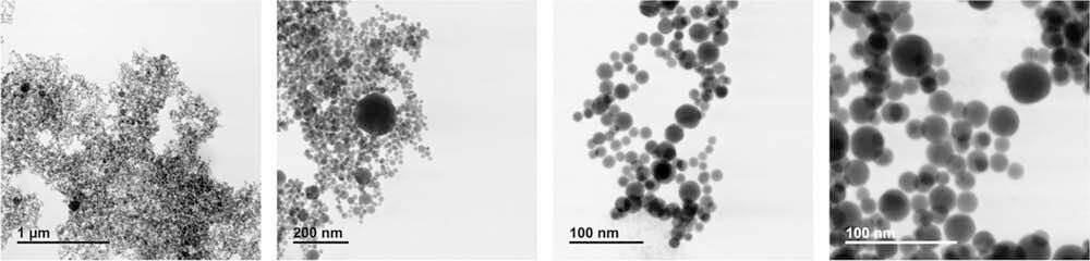
The study tracked both total silver and ionic silver after exposure to deionized water, moderately hard water, and concentrated artificial alveolar fluid. Mineral-rich media changed the appearance and dispersion state of the samples, so the authors combined bulk silver measurements with microscopy rather than treating a color change or a single sizing result as proof of dissolution.
ICP-OES measurements and ion-selective-electrode results were interpreted alongside ultracentrifugation and microscopy. The article reports low ion emission for EVQ-218 and emphasizes that mineral association, precipitation, and electrode interference must be distinguished from true particle dissolution.
Surface chemistry and particle fate
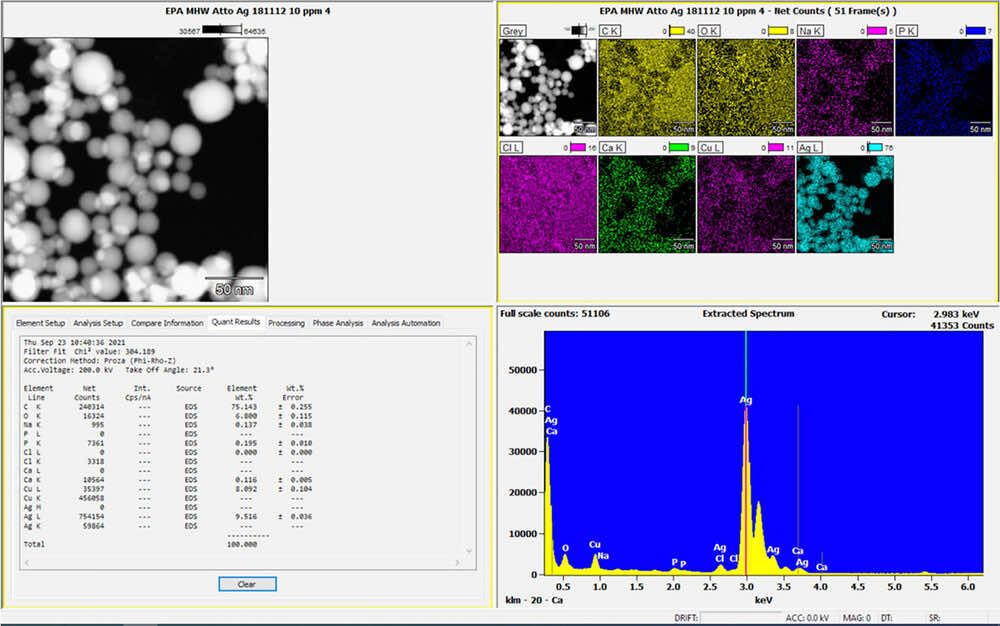
STEM/EDS mapping showed how calcium-, carbon-, oxygen-, and phosphorus-containing minerals associated with silver particles after media exposure. These maps let the authors distinguish an elemental silver signal from the surrounding mineral precipitate and compare the morphology retained by each material.
EELS analysis was used to examine the particle surface and compare EVQ-218 with the citrate-containing reference. The authors interpret the EVQ-218 spectra as evidence of a bare silver surface without an added stabilizing shell.
Long-term stability
The article includes microscopy of EVQ-218 material produced in 2007 and imaged in 2020. The authors report retained particle identity and uniform silver density over that interval. This observation supports long-term material stability in the stored sample; it does not by itself establish shelf life in every formulation, package, or application environment.
Conclusions and boundaries
The paper reports that EVQ-218 was comparable with the NIST-traceable reference in particle morphology and uniformity while differing in surface chemistry and emission behavior. Its central conclusion is that the laser-produced particles combine a narrow size distribution with a stable, surfactant-free silver surface.
These are physicochemical characterization results. Application performance, toxicology, environmental fate, and clinical outcomes require their own studies and should not be inferred from particle characterization alone.